
Sometimes atomic resolution imaging can be a big help in understanding how molecular machinery works. A news release from the Biodesign Institute of Arizona State University suggests that this may become easier to do “New method opens crystal clear views of biomolecules – Fundamental discovery triggers paradigm shift in crystallography“:
A scientific breakthrough gives researchers access to the blueprint of thousands of molecules of great relevance to medicine, energy and biology. In a new study, researchers from Arizona State University (ASU), Deutsches Elektronen-Synchrotron (DESY) and Stanford Linear Accelerator Laboratory (SLAC) describe a simple way to determine the 3-dimensional structures of proteins and other molecules, many of which are inaccessible by existing methods.
The research findings appear in the current issue of the journal Nature [abstract].
Imaging the molecular building blocks of living things at the atomic scale is tricky. Often, the most difficult step is getting such molecules to form high-quality crystals needed for X-ray imaging. This international team describes a new method that can produce sharp images relying on crystals with very small imperfections, using the world’s brightest X-ray source at the Department of Energy’s SLAC National Accelerator Laboratory.
“Once the full potential of the new method is understood, it could turn out to be one of the biggest advances since the birth of crystallography,” said Mike Dunne, director of the Linac Coherent Light Source (LCLS) X-ray laser, a DOE Office of Science User Facility.
The structures of biomolecules reveal their modes of action and provide insights into the workings of the machinery of life. Unlocking the molecular structure of particular proteins, for example, can provide the basis for developing tailor-made drugs against numerous diseases or advancing clean energy technologies with the efficiency of nature and the stability of engineered systems.
The chosen target of the new study is a critical enzyme known as photosystem II. Petra Fromme, co-author of the new study and director of the Biodesign Institute’s Center for Applied Structural Discovery (CASD) at ASU highlights the importance of this molecule:
“Life on earth relies directly on photosynthesis—the natural process of converting light energy to chemical energy in plants and algae. The critical first step is carried out by the photosystem II protein complex, which uses sulight [sic] to split abundant sources of water (H2O) into hydrogen, electrons and all the oxygen that sustains our planet,” she says.
In 2011,the team of researchers at ASU, (including Fromme and John Spence, professor of Physics, along with their collaborators), pioneered a technique called serial femtosecond crystallography. Here, structure determination is based on imaging of thousands of nanocrystals, so small that they can not even be seen under a microscope. The nanocrystals are hit “on-the-fly” by X-ray pulses that are so strong that they destroy any solid material but so short that a diffraction image is obtained before the destruction occurs.
The method was further developed in 2014, and was used to produce the first snapshot images of the water splitting process in photosysnthesis [sic]. It wasn’t clear however if the researchers would ever be able to see the dynamics of the process at atomic scale. Such fine detail coupled with dynamic imaging is essential for understanding the mechanisms of photosynthesis that split water and produce oxygen with visible light and earth abundant metals. Traditional X-ray crystallography methods could achive [sic] high resolution but suffer from damage from the X-rays and are not suitable to see dynamics of the process.
“This new method allows us to significantly improve the resolution of the images we took of photosystem II and gives us a new tool for capturing the dyanmics [sic] of some of life’s most important processes. When we understand how photosynthesis works at the chemical level we can use this as a blueprint for developing clean and renewable energy technologies,” Fromme adds.
“The best crystals are crystals with small imperfections”
More than 100 years ago, Australian-born British physicist William Lawrence Bragg found a way to use X-rays to probe the interior of crystals, which consist of regular arrays of atoms or molecules. This discovery launched the field of X-ray crystallography, one of the most important techniques for analyzing the structures of materials, chemical processes and biological molecules.
When X-rays pass through the crystal, they scatter off the protein molecules and form a pattern on a detector. This diffraction pattern is dominated by bright spots known as Bragg peaks, which researchers use to reconstruct the atomic structure of the molecules.
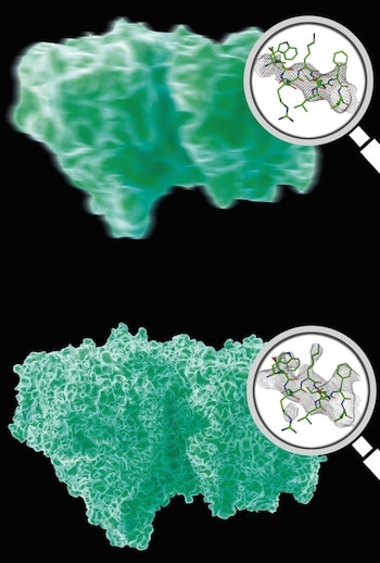
A perfectly ordered crystal would produce nothing but Bragg peaks. However, disorder limits the number of detectable peaks and therefore the resolution of the molecular image that can be obtained from the peaks alone.
The small displacements lead to “termination of the Bragg peaks”, but allow scientists to “see” the scattering from the sum of the individual molecules in the crystals. These tiny molecular displacements from a perfectly ordered crystal arrangement produce gently rippling patterns between and beyond the sharp Bragg peaks. While these patterns, known as “continuous diffraction,” have been actively studied, they had not been considered capable of producing high-resolution molecular images.
“We’ve now demonstrated that we can actually use the continuous diffraction of imperfect crystals to obtain better molecular images than with Bragg peaks alone,” said Kartik Ayyer, the study’s first author from the Center for Free-Electron Laser Science (CFEL) at the German research center DESY.
“Phased in”
Even when good diffraction is available the structure of a complex molecule is still extremely difficulty to determine. Without knowing the phase – the lag of the crests of one diffracted wave to another – it is not possible to compute an image of the molecule from the measured diffraction pattern.
To solve the tricky phase puzzle, more information must be known than just the intensity of the measured Bragg spots. Sometimes this information can be derived by X-[ray] analysis of crystals of chemically modified molecules, or inferred from the structure of a closely-related molecule, but these approaches are time consuming and can bias the results. Bragg peaks are strong, since they arise from constructive interference (like a crowd clapping in unison), but they are sparse and of limited content. The continuous diffraction used in the new method fills in the gaps in and beyond the Bragg peaks, giving vastly more information, which can be used to directly obtain the phase and improve the resolution.
ASU physicist and study co-author John Spence elaborates:
“In 1913, Paul Debye showed that Bragg diffraction from vibrating atoms in a crystal scatter between the Bragg spots. Henry Chapman (XFEL scientist with DESY and corresponding author) was able to show how, in a similar way, the non-Bragg scattering from LCLS X-ray snapshots from imperfect crystals can be used to provide a resolution-enhanced image of the protein molecule, and also solve the phase problem.
This simple concept leads to a paradigm shift in crystallography — the most ordered crystals are no longer the best to analyze. Imperfect crystals arise because some molecules do not align perfectly with majority of the molecules within the crystal [see Figure A in the original article], which creates a continuous diffraction pattern similar to those predicted for single molecules. Single molecule diffraction is the Holy Grail of modern X-ray science because it eliminates the crystallization process altogether, but has never been possible because the signals from individual molecules are far too weak.
“For the first time we have access to single molecule diffraction – we have never had this in crystallography before,” Chapman says. The technique elegantly weds X-ray diffraction of crystals and X-ray imaging of single particles, providing the best of both worlds.
The technique could provide a stepping-stone toward detailed imaging of single particles, including the large number of biological specimens that cannot easily be crystallized[.]
Fromme states that the new technique also opens the field for time resolved movies of biomolecules at work. “While perfect crystalline order restricts the movement of molecules, the new method allows us to study dynamic processes in single crystals of biomolecules.”
No doubt the earliest applications of this technology will focus on biomolecules and biomedical applications, as described above. As experience with the technology accumulates, however, it would be interesting to see it applied to dynamic artificial molecular machinery, such as this and these and these and this example of dynamic structural DNA nanotechnology.
—James Lewis, PhD