Molecular Labeling of Gold Nanowells Towards
Ultra-high Density 'On Tip' Fluorescence Analysis
Akito Ishida*
PRESTO JST Co. and Kyoto Prefectural University,
Kyoto 606-8522, JAPAN
This is an abstract
for a presentation given at the
Ninth
Foresight Conference on Molecular Nanotechnology.
There will be a link from here to the full article when it is
available on the web.
Surface plasmon (SP) is the charge density wave on a gold or silver surface excited by visible and near-IR light. The electromagnetic field is strongly localized on the surface and the propagation length is very long. Moreover, the field intensity is remarkably enhanced compared with that of the incident light. As an excitation source for photofunctional molecules attached on a gold or silver surface, these features offer distinct advantages over a conventional direct photoirradiation. [1] Recently, it has been reported that the SP field distribution can be controlled by holes with a sub-micron diameter prepared on the gold surface and the SP field can be strongly localized at the rims. [2] The regular array of such nano-sized gold holes prepared on a glass substrate may be utilized as vessels for fluorescence analysis. The spot density will be dramatically increased compared with a conventional multi-well plate and a DNA chip. In this study, selective molecular labeling of the "gold nanowells" and fluorescence imaging have been studied to realize the novel concept.
The nano-holes on a 100 nm gold film have been prepared by projection method [2] using polystyrene spheres with diameters of 200, 400, 500 nm or 1000 nm. After removal of the spheres, the gold surface and the bottom of the nanowells (i. e., exposed glass surface) were modified by a thiol derivative and an aminoalkylsilane derivative , respectively. The amino terminated bottom of the nanowells were then modified by amine-reactive fluorescence probes such as BODIPY TMR-X SE (Molecular Probes, Inc.). The fluorescence imaging has been carried out by a fluorescence microscope (OLYMPUS IX-70), a semiconductor laser (404, 532, or 635 nm), and a cooled-CCD camera (SPOT-JR). Figure 1 shows fluorescence image of the BODIPY TMR modified nanowells by graze illumination of 404 nm. The intense fluorescence demonstrates that the BODIPY molecules modified in the nanowells have been effectively excited. Fluorescence detection of antigen-antibody interaction and hybridization of DNA in the nanowells will be discussed.
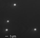
Figure 1
References
- A. Ishida, et al., Analyst, 535-540, 2000; Chem. Phys. Lett., 322, 242-246 (2000); Chem. Comm., 1299-1300, 1999; Nanotechnology, 10, 308-314 (1999) ; Chem. Commun., 57-58, 1998; Chem. Lett., 267-268, 1998.
- C. Soennichsen, et al., App. Phys. Lett., 76, (1999) 140.
Abstract in RTF format 7,735 bytes
*Corresponding Address:
Akito Ishida
PRESTO JST Co. and Kyoto Prefectural University
Dept. of Material Design, Kyoto Pref. Univ.
Shimogamo Hangi-cho 1-5, Sakyo-ku, Kyoto 606-8522, JAPAN
Phone: +81-75-703-5443
Fax: +81-75-703-5445
Email: [email protected]
|