
Researchers from UCLA’s California NanoSystems Institute and Northwestern University have combined multiple imaging techniques to produce high quality 3D images of platinum nanoparticles, allowing advanced visualization of atomic-scale structural defects (an important advancement over X-ray crystallography).
The original 2012 work, published in Nature and posted by Jim Lewis here, used electron tomography to study 10-nm gold particles and was described at Phys.org:
…
“This is the first experiment where we can directly see local structures in three dimensions at atomic-scale resolution — that’s never been done before,” said Jianwei (John) Miao, a professor of physics and astronomy and a researcher with the California NanoSystems Institute (CNSI) at UCLA.
…
X-ray crystallography is a powerful technique for revealing the structure of perfect crystals, which are materials with an unbroken honeycomb of perfectly spaced atoms lined up as neatly as books on a shelf. Yet most structures existing in nature are non-crystalline, with structures far less ordered than their crystalline counterparts — picture a rock concert mosh pit rather than soldiers on parade.
…
Miao and his colleagues used a scanning transmission electron microscope to sweep a narrow beam of high-energy electrons over a tiny gold particle only 10 nanometers in diameter (almost 1,000 times smaller than a red blood cell). The nanoparticle contained tens of thousands of individual gold atoms, each about a million times smaller than the width of a human hair. These atoms interact with the electrons passing through the sample, casting shadows that hold information about the nanoparticle’s interior structure onto a detector below the microscope.Miao’s team discovered that by taking measurements at 69 different angles, they could combine the data gleaned from each individual shadow into a 3-D reconstruction of the interior of the nanoparticle. Using this method, which is known as electron tomography, Miao’s team was able to directly see individual atoms and how they were positioned inside the specific gold nanoparticle.
…
The new study, using multiple imaging techniques, will be published in an upcoming issue of Nature, and includes a video showing three-dimensional volume renderings (available for viewing at Phys.org:
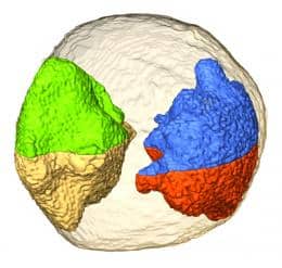
The authors describe being able to see how the atoms of a platinum nanoparticle—only 10 namometers in diameter—are arranged in three dimensions. They also identify how the atoms are arranged around defects in the platinum nanoparticle.
…
This novel method is a combination of three techniques: scanning transmission electron microscopy, equally sloped tomography (EST) and three-dimensional Fourier filtering. Compared to conventional CT, the combined method produces much higher quality 3-D images and allows the direct visualization of atoms inside the platinum nanoparticle in three dimensions.
…
“This is the first instance where the three-dimensional structure of dislocations in nanoparticles has been directly revealed at atomic resolution,” Ajayan said. “The elegant work demonstrates the power of electron tomography and leads to possibilities of directly correlating the structure of nanoparticles to properties, all in full 3-D view.” Defects can influence many properties of materials, and a technique for visualizing these structures at atomic resolution could lead to new insights beneficial to researchers in a wide range of fields.
-Posted by Stephanie C