Two research teams present two different methods for using single strands of DNA to link various nanoparticles into complex 3D arrays: one using DNA hairpins for dynamic reconfiguration and the other using a DNA origami scaffold.
Two research teams present two different methods for using single strands of DNA to link various nanoparticles into complex 3D arrays: one using DNA hairpins for dynamic reconfiguration and the other using a DNA origami scaffold.
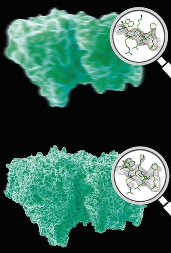
A paradigm shift in analyzing diffraction from smaller, less perfect crystals yields improved resolution and enables directly determining the phase of the diffraction pattern.
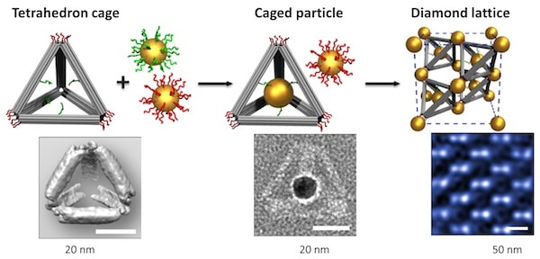
Encapsulating enzymes in nanocages engineered using structural DNA nanotechnology increases enzymatic digestion and protects enzymes from degradation.
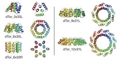
New families of protein structures, barrel proteins for positioning small molecules, self-assembling protein arrays, and precision sculpting of protein architectures highlight de novo protein design advances.
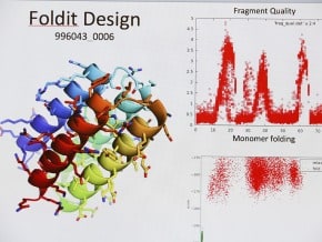
Computational design of proteins satisfying predetermined geometric constraints produced stable proteins with the designed structure that are not found in nature.
A fully automated design protocol generates dozens of designs for proteins based on helix-loop-helix-loop repeat units that are very stable, have crystal structures that match the design, have very different overall shapes, and are unrelated to any natural protein.
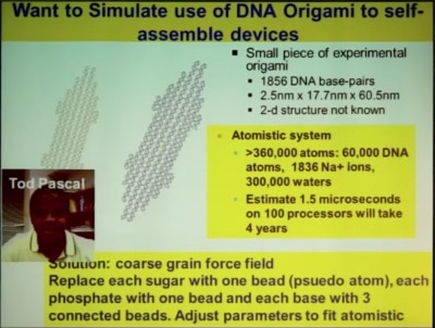
Prof. William Goddard presented four advances from his research group that enable going from first principles quantum mechanics calculations to realistic nanosystems of interest with millions or billions of atoms.
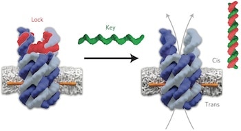
DNA building blocks mimic biological ion channels to more precisely control which molecules can cross a biological membrane.
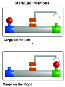
A molecular robotic arm synthesized from small synthetic organic molecules uses cyclic changes in pH and other reaction conditions to grab and release a cargo molecule, and swing the cargo back and forth between the two ends of the molecular platform.
The positions of 3769 tungsten atoms in a tungsten needle segment were determined to a precision of 19 pm (0.019 nm), including the position of a single atom defect in the interior of the sample, by using aberration-corrected scanning transmission electron microscopy and computerized tomography.